Which of the Following Indicates Possible Ms During an Mri
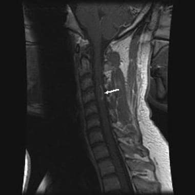
Magnetic resonance imaging MRI plays a crucial role in multiple sclerosis MS diagnosis disease monitoring prognostication and research. The spinal cord may or may not be focally enlarged.
Which Findings On Mri Suggest Multiple Sclerosis Ms
According to the findings 493 of MS patients who received injections had spots of signal intensity in their magnetic resonance imaging MRI scans which is a sign of gadolinium build up.
. A client is scheduled for magnetic resonance imaging MRI. 3 MULTIPLE CHOICE OPTIONS. 1 Other causes of white spots on a brain MRI include.
During the client teaching what will the nurse discuss. Its thought to be the result of an immune system attack. How much air is inhaled and exhaled and how quickly it is exhaled.
Multiple Sclerosis is a potentially disabling disease of the _____. Several important practice guidelines updates for MRI in MS have been published recently including the 2017 revised McDonalds Criteria 1 Magnetic Resonance Imaging in MS network guidelines 2. 1 MS diagnosis requires demonstration of disease dissemination in space DIS and time DIT and exclusion of other conditions that can mimic.
Enlargement of the cord is usually seen with active disease. The spirometry pulmonary function test measures _____. Hi XXXX Thanks for being in follow-up again.
I should make it clear to you that ALS is not a disease which is diagnosed on MRIIn factMRI of brain does not have even a bit of role in diagnosing ALS. Multivariable prediction model of lesion characteristics differentiating asymptomatic PML from new MS lesions. Unilateral muscle weakness is a neurological abnormal clinical sign that indicates a nervous system malfunction but not which specific causation and the cause might be with the spine and not brain so IF you havent as yet had a spinal MRI as well as blood peripheral nerve tests etc etc it is far to early in your diagnostic work up to point towards or away from anything yet.
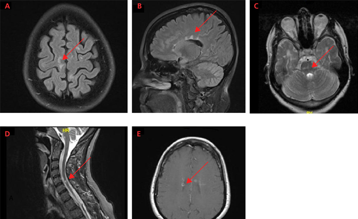
Which of the following indicates possible MS during an MRI. However the relatively low sensitivity of this test can make a definite diagnosis of nmo or nmo spectrum disorder more. The follow-up MRI shows new focal periventricular MS lesions in the corpus callosum C and D closed arrowheads.
Brain stem magnetic resonance imaging and evoked potential studies of symptomatic multiple sclerosis patients. Silent strokes often occur in deeper regions of the brain and are usually caused by blockage of small blood vessels. 29 30 the diagnosis of nmo can be supported by antibody findings against aquaporin 4.
Getty Images Nearly everyone with multiple sclerosis MS has signs of lesions in the brain as shown by magnetic resonance. Mills RJ Young CA Smith ET. T-1 weighted with gadolinium- may show bright areas enhancing lesions that indicate areas of active inflammation.
Comi G Filippi M Martinelli V Set al. MS causes demyelination or the damage of myelin. 32 years old male had a brain MRI at the age 30 what prompt me to get it was constant bad headaches and a not very good short term memoryI had braces at the time so the imaging wasnt very clear however in the report it said there are at least six scattered foci of the juxtacortical white matter hyperintensity invoking the left acceptable left frontal parietal and.
2 A brain tumor such as lymphoma B12 deficiency Infection such as Lyme disease or HIV Lupus Migraines Multiple sclerosis MS Risk Factors. 3 MULTIPLE CHOICE OPTIONS. 1 Relapsing-remitting MS MOST COMMON 2 Secondary progressive MS second most common 3 Primary progressive MS 4 Relapsing-progressive MS not very common.
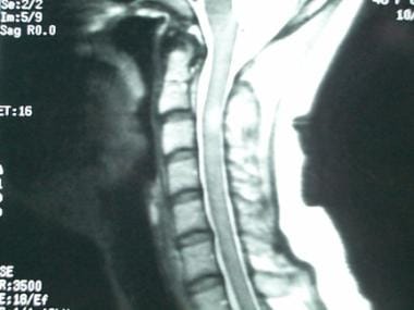
The most common condition that mimics ms lesions is neuromyelitis optica nmo which can present with brain lesions in up to 70 of cases. Furthera clinical history comprising only of fasciculationsis not. MS lesions on the spinal cord can be seen on an MRI scan as shown here.
The examination will take only 15 minutes Do you experience any claustrophobia You must be NPO for the day before the examination You must remove all jewelry but can wear your wedding ring A client undergoing a diagnostic examination for. Central trigeminal involvement in multiple sclerosis using high-resolution MRI at 3T. Most MS plaques appear hyperintense on T2-weighted images.
Larger active lesions may. Common MRI sequences used in MS include. MRI with contrast dye can indicate MS disease activity by showing a pattern consistent with inflammation of active demyelinating lesions.
MRI is not the investigation for ALS. These types of lesions are new or getting bigger due to. T-1 weighted without gadolinium- may show dark areas hypointensities that are thought to indicate areas of permanent nerve damage.
The sensitivity of T 1 - and T 2-weighted MRI to white matter WM lesions and to regional as well as global atrophy has made this modality central to the diagnosis and treatment monitoring of multiple sclerosis MS1-4 Paradoxically these findings correlate only weakly with clinical disability an incongruity due MRIs insensitivity to microscopic pathology and lack of specificity. Magnetic resonance imaging MRI was formally included in the diagnostic work-up of patients presenting with a clinically isolated syndrome CIS suggestive of multiple sclerosis MS in 2001 by an International Panel of experts. Multiple sclerosis MS is a chronic disease of your central nervous system CNS.
Multiple Sclerosis
Which Findings On Mri Suggest Multiple Sclerosis Ms
2
No comments for "Which of the Following Indicates Possible Ms During an Mri"
Post a Comment